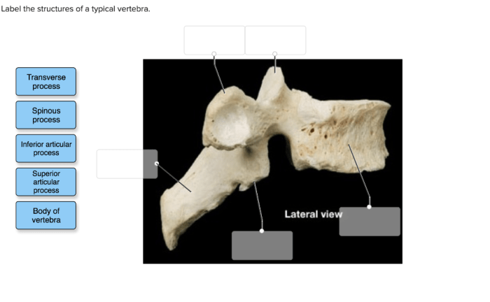
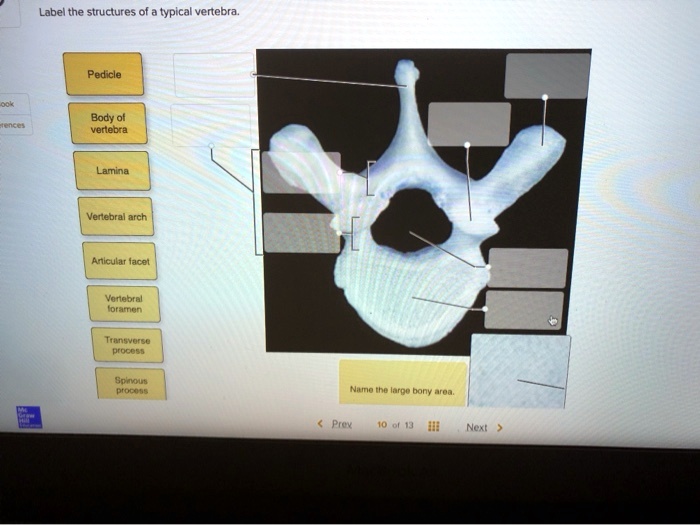
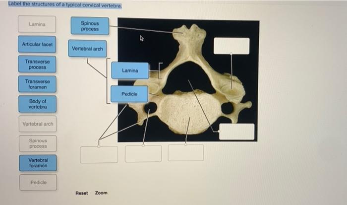
Label the structures of the typical vertebra – Embarking on a journey into the realm of human anatomy, we delve into the intricacies of the typical vertebra, the building blocks of our resilient spinal column. This comprehensive guide will meticulously dissect each structure, unraveling its form, function, and significance in maintaining our posture and facilitating movement.
From the robust vertebral body that bears the weight of our bodies to the intricate vertebral arch that encloses the delicate spinal cord, each component plays a vital role in the harmonious functioning of our skeletal system.
1. Vertebral Body
The vertebral body, also known as the centrum, is the main, weight-bearing component of a vertebra. It is located anteriorly and is shaped like a cylinder or a spool. The vertebral body is responsible for supporting the weight of the body and distributing it evenly across the spine.
Vertebral Body Comparison
| Region | Shape | Size | Function |
|---|---|---|---|
| Cervical | Small and rounded | Thinner | Supports the head and allows for greater mobility |
| Thoracic | Larger and heart-shaped | Thicker | Supports the rib cage and provides attachment for the ribs |
| Lumbar | Largest and kidney-shaped | Thickest | Supports the weight of the upper body and provides stability |
2. Vertebral Arch
The vertebral arch is a posterior structure that extends from the vertebral body and encloses the spinal canal. It consists of two pedicles and two laminae. The pedicles are short, thick columns that connect the vertebral body to the laminae.
The laminae are broad, flat plates that form the roof and sides of the vertebral arch. The vertebral arch helps to protect the spinal cord and provides attachment for muscles and ligaments.
Diagram of Vertebral Arch
[Diagram of vertebral arch with clear labels]
3. Transverse Processes

The transverse processes are two lateral projections that extend from the junction of the pedicles and laminae. They are oriented horizontally and provide attachment for muscles and ligaments. The transverse processes also help to stabilize the spine and limit lateral movement.
Table of Muscles and Ligaments Attached to Transverse Processes
| Muscle/Ligament | Attachment |
|---|---|
| Multifidus muscle | Medial surface |
| Rotatores muscle | Medial surface |
| Interspinalis ligament | Between adjacent laminae |
| Supraspinal ligament | Between the spinous processes |
4. Spinous Process

The spinous process is a single, midline projection that extends posteriorly from the junction of the laminae. It is shaped like a triangle or a spine and provides attachment for muscles and ligaments. The spinous process also helps to protect the spinal cord and limit flexion and extension of the spine.
Illustration of Spinous Process in Muscle Action
[Illustration demonstrating the role of the spinous process in muscle attachment and action]
5. Superior and Inferior Articular Processes
The superior and inferior articular processes are four projections located on the posterior aspect of the vertebral arch. The superior articular processes are located on the upper surface of the pedicles and face upward. The inferior articular processes are located on the lower surface of the laminae and face downward.
The articular processes form facet joints with the adjacent vertebrae, allowing for movement of the spine.
Articular Process Comparison
| Region | Orientation | Function |
|---|---|---|
| Cervical | Flat and oval | Allow for greater range of motion, including flexion, extension, and rotation |
| Thoracic | Round and interlocking | Restrict movement to mainly flexion and extension |
| Lumbar | Large and cylindrical | Allow for lateral bending and some rotation |
6. Foramen Magnum

The foramen magnum is a large opening located on the posterior surface of the occipital bone, which is the first vertebra. It is the opening through which the spinal cord enters the skull. The foramen magnum is bounded by the occipital condyles, which are two projections that articulate with the atlas, the first cervical vertebra.
Illustration of Foramen Magnum
[Illustration or diagram of the foramen magnum in relation to the other structures of the vertebra]
7. Vertebral Canal
The vertebral canal is a hollow space that runs through the center of the vertebral arch. It is formed by the vertebral body anteriorly and the vertebral arch posteriorly. The vertebral canal contains and protects the spinal cord.
Cross-Sectional Diagram of Vertebral Canal
[Cross-sectional diagram of the vertebral canal showing the surrounding structures]
8. Intervertebral Disc
The intervertebral disc is a fibrocartilaginous structure located between adjacent vertebrae. It is composed of a tough outer layer called the annulus fibrosus and a soft, gelatinous inner layer called the nucleus pulposus. The intervertebral disc provides cushioning and flexibility to the spine, allowing for movement and shock absorption.
Illustration of Intervertebral Disc, Label the structures of the typical vertebra
[Illustration or diagram of the intervertebral disc in relation to the adjacent vertebrae]
FAQ Resource: Label The Structures Of The Typical Vertebra
What is the function of the vertebral body?
The vertebral body bears the weight of the body and provides structural support to the spine.
What is the vertebral arch?
The vertebral arch forms the posterior portion of the vertebra and encloses the spinal canal, which houses the spinal cord.
What is the role of the transverse processes?
The transverse processes provide attachment points for muscles and ligaments, contributing to the stability and flexibility of the spine.